
A groundbreaking convergence of artificial intelligence and medical imaging is fundamentally transforming how physicians detect and diagnose cancer, with new research demonstrating unprecedented accuracy rates that could revolutionize early intervention strategies. The integration of deep learning algorithms with advanced imaging technologies represents not merely an incremental improvement but a paradigmatic shift in oncological diagnostics, promising to address long-standing challenges in sensitivity, specificity, and accessibility that have plagued traditional screening methods.
According to recent findings published in Nature Communications Medicine, researchers have developed sophisticated AI models capable of detecting subtle cancerous lesions that frequently elude human observers, even experienced radiologists. The study demonstrates that machine learning systems trained on vast datasets of medical images can identify patterns and anomalies with remarkable consistency, achieving detection rates that surpass conventional diagnostic protocols. This technological advancement arrives at a critical juncture, as healthcare systems worldwide grapple with increasing cancer incidence rates and the imperative to diagnose malignancies at earlier, more treatable stages.
The implications extend far beyond academic research laboratories. Major healthcare institutions are already implementing AI-assisted diagnostic tools in clinical settings, fundamentally altering workflows and decision-making processes. These systems function not as replacements for human expertise but as powerful augmentation tools, providing radiologists with additional analytical capabilities and reducing the cognitive burden associated with reviewing thousands of images. The technology’s capacity to process and analyze imaging data at scales impossible for human practitioners represents a quantum leap in diagnostic efficiency.
The Architecture of Intelligence: How Deep Learning Decodes Medical Images
The technical foundation underlying these AI diagnostic systems relies on convolutional neural networks, a class of deep learning algorithms specifically designed to process visual information. These networks undergo extensive training phases, analyzing millions of annotated medical images to develop pattern recognition capabilities. The Nature Communications Medicine research details how these algorithms learn to distinguish between benign and malignant tissue characteristics by identifying subtle variations in density, texture, and morphology that may indicate cancerous transformation.
What distinguishes contemporary AI diagnostic systems from earlier computer-aided detection tools is their ability to learn hierarchical representations of image features. Rather than relying on predefined rules or simple pattern matching, modern deep learning models develop increasingly sophisticated internal representations through multiple processing layers. This architectural approach enables the systems to capture both low-level details—such as pixel-level intensity variations—and high-level conceptual features that correlate with specific pathological conditions. The result is a diagnostic capability that approaches and, in some cases, exceeds human-level performance across various cancer types.
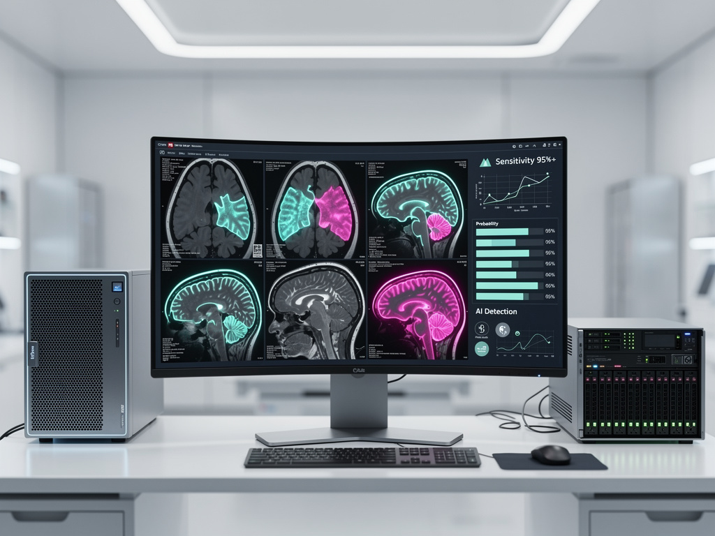
Validation and Performance Metrics: Measuring Diagnostic Superiority
The validation of AI diagnostic systems requires rigorous testing protocols that assess performance across diverse patient populations and imaging modalities. The Nature Communications Medicine study employed extensive datasets encompassing thousands of cases, with performance evaluated using standard metrics including sensitivity, specificity, and area under the receiver operating characteristic curve. Results demonstrated that the AI models achieved sensitivity rates exceeding 95% for certain cancer types, with false positive rates significantly lower than those associated with traditional screening approaches.
These performance improvements translate directly into clinical benefits. Higher sensitivity means fewer missed diagnoses, enabling earlier therapeutic intervention when treatment outcomes are most favorable. Reduced false positive rates minimize unnecessary follow-up procedures, biopsies, and the psychological distress associated with false alarms. The economic implications are equally substantial, as more accurate initial diagnostics can reduce healthcare costs associated with redundant testing and delayed treatment of advanced-stage cancers.
Implementation Challenges and Regulatory Considerations
Despite impressive technical achievements, the pathway to widespread clinical adoption confronts significant obstacles. Regulatory frameworks governing medical devices and diagnostic tools must evolve to accommodate AI-based systems whose performance characteristics differ fundamentally from traditional diagnostic equipment. The U.S. Food and Drug Administration and international regulatory bodies are developing new evaluation criteria specifically tailored to machine learning algorithms, recognizing that these systems require ongoing monitoring and potential retraining as they encounter new data.
Integration into existing clinical workflows presents additional challenges. Healthcare institutions must invest in computational infrastructure capable of supporting AI applications, including high-performance computing resources and secure data storage systems. Radiologists and other healthcare professionals require training to effectively utilize AI-assisted diagnostic tools, understanding both their capabilities and limitations. The human-machine interface design becomes critical, as systems must present information in formats that facilitate rapid comprehension and support clinical decision-making without introducing new sources of error or confusion.
Equity and Access: Democratizing Advanced Diagnostics
One of the most compelling promises of AI-powered diagnostic systems lies in their potential to democratize access to high-quality cancer screening, particularly in underserved regions lacking specialized radiological expertise. Once developed and validated, AI models can be deployed at relatively low marginal cost, enabling community hospitals and rural healthcare facilities to offer diagnostic capabilities previously available only at major academic medical centers. This democratization could address persistent healthcare disparities that contribute to differential cancer outcomes across socioeconomic and geographic divides.
However, realizing this potential requires deliberate attention to algorithmic fairness and generalizability. AI models trained predominantly on data from specific demographic groups may exhibit reduced performance when applied to populations with different characteristics. Researchers are increasingly focused on developing training strategies that ensure robust performance across diverse patient populations, incorporating data from multiple geographic regions, ethnic backgrounds, and healthcare settings. The Nature Communications Medicine research emphasizes the importance of diverse training datasets in achieving equitable diagnostic performance.
The Economic Calculus: Cost-Benefit Analysis of AI Diagnostics
The economic case for AI-assisted cancer diagnostics extends beyond direct cost savings from reduced false positives and earlier detection. Healthcare systems face mounting pressure to manage increasing imaging volumes while maintaining diagnostic quality—a challenge exacerbated by radiologist shortages in many regions. AI systems can process routine cases efficiently, enabling human specialists to focus attention on complex or ambiguous cases requiring nuanced clinical judgment. This division of labor optimizes resource utilization and potentially extends the effective capacity of existing radiological workforces.
Initial implementation costs, including software licensing, infrastructure upgrades, and training programs, represent significant investments that healthcare institutions must carefully evaluate. However, longitudinal analyses suggest that these costs are offset by improved diagnostic accuracy, reduced litigation risk associated with missed diagnoses, and enhanced patient throughput. As AI diagnostic tools mature and become more widely adopted, economies of scale will likely drive down costs, making these technologies increasingly accessible to healthcare organizations of varying sizes and resource levels.
Future Trajectories: Beyond Detection to Prediction
The evolution of AI in oncological imaging is progressing beyond simple detection toward predictive analytics and treatment planning. Emerging research explores how machine learning algorithms can analyze imaging data to predict tumor behavior, treatment response, and patient outcomes. These predictive capabilities could enable truly personalized medicine, with treatment strategies tailored to individual patient characteristics and tumor biology as revealed through advanced image analysis.
Integration with other data modalities represents another frontier. Combining imaging data with genomic information, electronic health records, and molecular biomarkers through multimodal AI systems could provide comprehensive patient assessments that inform both diagnostic and therapeutic decisions. The Nature Communications Medicine research points toward this integrative future, suggesting that the full potential of AI in oncology will be realized through systems that synthesize diverse information sources into actionable clinical insights.
The Human Element: Redefining Radiological Practice
As AI systems assume greater responsibility for routine diagnostic tasks, the role of radiologists is evolving rather than diminishing. The profession is shifting toward more consultative functions, with specialists focusing on complex case interpretation, quality assurance of AI outputs, and direct patient interaction. This evolution requires changes in medical education and training programs, ensuring that future radiologists develop expertise in both traditional image interpretation and the effective utilization of AI tools.
The collaborative relationship between human expertise and machine intelligence represents the optimal paradigm for cancer diagnostics. AI systems excel at consistent, high-volume analysis and pattern recognition across vast datasets, while human practitioners contribute contextual understanding, clinical judgment, and the ability to integrate diverse information sources. This synergy, rather than competition between human and artificial intelligence, offers the most promising path toward improved cancer detection and patient outcomes. The transformation underway in medical imaging exemplifies how artificial intelligence, properly developed and implemented, can augment human capabilities and advance the fundamental mission of medicine: improving human health and alleviating suffering.

